Friday, July 29, 2016

1. Study Objective

H0

H1


We needed to collect data.... (to be continued)

2. Variables 

2.1 Collected Variables


Gender
Age
Weight
Height
Waist circumference
Hip circumference
Distance, d, between the iliac crest and the umbilicus

2.2 Calculated Variables 


Body mass index (BMI)

Waist-to-hip ratio

3. Recruitment


We recruited 56 random participants, of which 30 were males and 26 were female, from the ages of 16 to 29. 


4. Privacy Measures


4.1 A full explanation of our study objective, study requirements and data collection procedure were given to every participant before commencement.


4.2 Participants were only allowed into the room one at a time. 


4.3 Chaperones of both genders were present during the entire duration of our data collection. 


4.4 The participants' names were not recorded. 


4.5 There was no removal of clothing except for the participants' shoes. 


4.6 Permission for the palpation of the iliac crests were granted by participants. 


4.7 The study data of every participant was kept confidential and all information were only used for our study and presentation purposes. 


5. Location and Duration

We held our data collection at a library room. The data collection roughly took about 4 hours in total, and roughly 3 minutes on each participant. 

6. Equipment

Digital weighing scale
Construction measuring rape
Tailoring measuring tape
Ruler
Paper
Masking tape
Sturdy file
Sleeping bag

7. Methodology

7.1 Study Organisation

We divided our data collection into 4 stations. The participants were required to go through the 4 stations in chronological order. 

Station 1: Gender and age recording
Station 2: Weight and height measurement
Station 3: Waist and hip circumference measurement 
Station 4measurement in a supine (lying down) position

7.2 Room Preparation 

Before the data collection started, we prepared the equipment needed for each station. 

Station 1: We prepared small pieces of paper for each participant to write down their gender and age.


Station 2: We brought along a digital weighing scale that was tared before each weight measurement. Using masking tapes and the construction measuring tape, we set up a height measurement scale. The height measurement scale was thoroughly checked to be perpendicular, with the 0 cm marking corresponding to the floor. A sturdy file was used to measure the participants' height. 


Station 3: Tailoring measuring tapes were used for the measurement of the participants' waist and hip circumferences. 


Station 4: A sleeping bag was spread out on the floor, so that participants would not have to lie directly on the floor. 

7.3 Data Collection Procedure

After the preparation of each stations, and following the privacy measures (mentioned in section 5), we called in our participant. 


Station 1: The participant was asked to fill in only his/her gender and age (as of that date) on a slip of paper. 


Station 2: Here the participant was measured for his/her weight and height. 


The participant's shoes had to be removed for this station. The participant was told to step on the weighing scale. Next, the participant was told to have their back against the wall, with their weight distributed equally over both feet, face looking straight ahead. A sturdy file was placed over the top of the participant's head, and checked to be parallel to the top of his/her head and the floor before the height measurement was recorded.

Station 3: Next, the participant wer
e measured for their waist and hip circumferences. 


The waist circumference was taken to be the circumference around the umbilicus, while the hip circumference was taken to be around the thickest part of the buttocks. We asked the participant to point out where his/her belly button to ascertain the waist circumference.  

Station 4: Lastly, participant were measured for d in a supine position. 


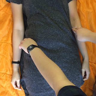
The participant was asked to lie down on the sleeping bag placed on the floor. With the participant's permission, we palpated for his/her iliac crests on both sides. We then placed a measuring tape across the level of the participant's iliac crests. Next, we asked the participant to point out their umbilicus. Using a ruler, we measured the distance, d, from the centre of the umbilicus to the level of the iliac crest.

8. Data Processing

With all the collected data, we proceeded to key them into SPSS. Our data findings would be continued in the next section (which tab name)....

Thursday, July 28, 2016

new intro

The iliac crest in Radiography


The iliac crest is an bony structure of the human body that can be externally palpated. It is felt at both sides of the lower abdomen, and extends towards the back of the body. Anatomically, it is the upper border of the hip bone. The iliac crests are major surface landmarks that allow us to achieve accurate positioning for many common radiographic examinations such as: 


- Abdomen/ Kidney-ureter-bladder examinations

- Pelvic examinations

- Lumbosacral spine examinations


However, the iliac crests are not always easily palpable. Patients who are obese, or have congenital or acquired deformities of the hip bone, usually present difficulties during palpation. But such conditions are not at all exclusive; different ethnic body types and different responses to physical touch can also create difficulties in palpating for the iliac crests. We have observed that there were patients who had been incredibly ticklish, and have seen many who were in pain or uncomfortable at the idea of being touched at their sides. 


Thus, it is not uncommon to see radiographers using the level of umbilicus ("belly button") as an alternative way to determine the level of the iliac crests, in actual practice. 

Vertebral location of iliac crest and the umbilicus


Transverse planes of the torso ("level") can be measured according to the 24 vertebrae levels. According to literature, the iliac crests are relatively fixed at the level of the fourth to fifth lumbar vertebrae (L4-L5)1. The level of the umbilicus, on the other hand, is not as precisely stated as that of the iliac crests, but is given to be between the L3 and L4 vertebral bodies2, with a normal variation ranging between L3 to L53. Based on a 2016 study3, of which 72.5% of the sample size were of Caucasian ethnicity, the level of the umbilicus was shown to be at a range of distances in relation to level of the iliac crests. 


Figure 1

This raises the question of whether the level of the umbilicus is an accurate measure of the level of the iliac crests. Our study aims to investigate its statistical reliability and correlation to patient size. 


1Bogduk, Nikolai; Endres, Stephen M. (2005). Clinical anatomy of the lumbar spine and sacrum (4th ed.). Elsevier Health Sciences. p. 106.

Chakraverty, R., Pynsent, P., & Isaacs, K. (2007). Which spinal levels are identified by palpation of the iliac crests and the posterior superior iliac spines? Retrieved July 28, 2016, from http://www.ncbi.nlm.nih.gov/pmc/articles/PMC2100271/

2Ellis, Harold (2006). Clinical Anatomy: Applied Anatomy for Students and Junior Doctors. New York: Wiley.

O'Rahilly, Ronan; Müller, Fabiola; Carpenter, Stanley; Swenson, Rand (2004). "Abdominal walls". Basic Human Anatomy: A Regional Study of Human Structure. Dartmouth Medical School.

3Evidence-Based Regulations : Annals of Plastic Surgery. (n.d.). Retrieved July 28, 2016, from http://journals.lww.com/annalsplasticsurgery/pages/articleviewer.aspx?year=2016


Figure 1: http://www.sbadandrmc.com/our-services-patient-education/digital-x-rays-70ea/abdominal-x-ray-kub/ (image) 

Thursday, July 21, 2016

Introduction

As radiographers-to-be, we come across problems in positioning some patients for certain radiographic procedures occasionally. Instead of choosing to do our Statistics Project on topics unrelated to our profession that we did consider earlier, we decided to do our project on something that can potentially impact on how we can position patients more efficiently and accurately.

As we racked our brains to think of what our topic should be on, we tried to observe more carefully during our clinicals to keep a lookout on what we can make our study on. We observed that some of the radiographers in the hospitals we were interning at were using a surface landmark that we were not taught in school - the umbilicus, also known as the belly button or navel.

A few radiographers we observed were using the level of the umbilicus as where the level of the iliac crest would be, while others took the level of the umbilicus to be a few centimetres away from the level of the iliac crest.

The iliac crest is a bony palpable landmark that forms the upper border of our pelvic bone. During certain radiographic procedures, radiographers are required to use this surface landmark to aid in positioning the patients correctly. Examples are for X-rays of the lumbar spine, hip and abdomen. However, this landmark may be quite difficult to palpate in some patients, either due to their abundant soft tissues, making it tough to feel for the bone, or because patients may be very ticklish in that area. 

We hence thought of doing a study on whether the umbilicus can be a good and reliable indicator of where the iliac crest is, and find out whether its reliability differs with the patient's size. This is for us to decide whether or not we can use it in place of the iliac crest, and for what kinds of patients we can use this landmark for, so that we can position patients with greater ease for the aforementioned procedures.

Our null hypothesis Ho:
The umbilicus is at the level of the iliac crest for people with different body mass index.

Our research hypothesis H1:
The umbilicus is not at the level of the iliac crest for people with different body mass index.

Dependent variable:
Distance between the level of the umbilicus and the iliac crest.

Independent variables:
Height, weight, body mass index
Waist circumference (measured at the level of the umbilicus)
Hip circumference (thickest part of the buttock area)
Waist-hip ratio

Tuesday, July 19, 2016

Literature review

According to most textbooks of human anatomy, the iliac crest is at the upper border of the level of the fourth lumbar vertebral body (L4) and this is a relatively fixed and standard position (Bogduk, 2005).

However, the level of the umbilicus is not as precisely stated as that of the iliac crest. This is as textbooks state that the umbilicus lies typically at a vertical level between the L3 and L4 vertebral bodies (Ellis, 2006), with a normal variation ranging between L3 to L5 vertebrae (O'Rahilly, 2004).
In old age, the umbilicus level goes down to a lower level due to reduced abdominal muscle tone (Singh, 2014).

We were not able to find literature on how body size would affect the level of the umbilicus.


References:

Bogduk, Nikolai; Endres, Stephen M. (2005). Clinical anatomy of the lumbar spine and sacrum (4th ed.). Elsevier Health Sciences. p. 106.

Ellis, Harold (2006). Clinical Anatomy: Applied Anatomy for Students and Junior Doctors. New York: Wiley.

O'Rahilly, Ronan; Müller, Fabiola; Carpenter, Stanley; Swenson, Rand (2004). "Abdominal walls". Basic Human Anatomy: A Regional Study of Human Structure. Dartmouth Medical School.

Singh (2014). Textbook of Anatomy Abdomen and Lower Limb; Volume 2. (2nd ed.) Elsevier Health Sciences. p. 299.

Monday, July 18, 2016

Methodology

With the aim of collecting as many data samples as possible, we booked a project room in the library and set up the room with stations, each to complete various measurements. There were 4 stations in total.

At the first station, participants were made to fill in their age and gender on a slip of paper.

Next, they were brought to the height and weight measuring station. They were asked to remove their shoes for this station for more accurate measurements. Their weight was taken by a digital weighing scale that was tared before they stepped on it for the measurement. Their height was taken manually using a measuring tape pasted against the wall. The measuring tape was checked to be perpendicular to the ground, with zero cm level corresponding to the floor. Each participant was told to have his or her back against the wall, weight distributed equally over both feet, face looking straight ahead. A box file was placed over the top of the participant's head, and checked to be parallel to the top of his/her head and the floor before the height measurement was recorded.

The third station was for their waist and hip circumference to be measured. This was done using a measuring tape. Their waist was taken to be around the umbilicus, while the hip was taken to be around the thickest part of the buttocks. We asked the participant to point out where their belly button was before placing the measuring tape around it for the waist measurement.

Finally, the participant was asked to lie down on the sleeping bag placed on the floor. We asked for permission to feel for their hip bone before palpating for their iliac crest on both sides and placing the measuring tape across to be at the same level. We then asked the participant to point out the level of their belly button again. Upon doing so, we measured using a ruler, the distance from the centre of the umbilicus, to the level of the iliac crest.

Each data statistic was carefully taken to minimise collection errors. No data was rejected. 

We recruited the help of participants who could each kindly spare us around five minutes of their time to help us with our data collection. They were students of Nanyang Polytechnic between the age range of 17 to 29. We tried to get an approximately equal mix of males and females in a spectrum of body sizes.